Wednesday, August 02, 2006
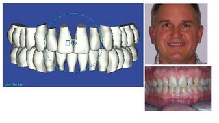
The accurate 3-D images of Dr. Snyder's teeth are digitally scanned photos made possible by Suresmile.

These are Dr. Snyder’s teeth before treatment. Note that the midlines are off and his upper front teeth are small with spaces.

His models are made and his braces are ready to be put on. Note that when we made plaster models to evaluate his bite there is more to the problem than meets the eye…his lower jaw can assume more than one position where the side teeth do not fit very well in this position….this multiple jaw position permitted the drift of the midlines, the shift of the midline, and contributed to the spacing in the upper.
Note that the lower front teeth are also quite worn due to this bite issue.

Again there is more present than meets the eye…note how small the lateral incisor truly is and the rotation of the premolar due to the bite permitting tooth drift over time.

The wear on the lower teeth is likely due to his strong eastern European facial type…strong chin/strong lower jaw. With strong lower jaw and a bite jaw permitted to assume multiple positions he doesn’t have a true home for his bite, especially if he grinds or clenches teeth at night. With strong jaws and bite without a true home the tiny lower teeth can get hammered over time and even wear down to little nubs.

The SureSmile software permits this view which superimposed the exact 3-D replica of his teeth over the side X-Ray. This shows the other important problem as to why he is straightening his teeth….his strong lower jaw is due to genetics and in his case this facial type also has strong lip muscles…these lip muscles are a tight band in front of his upper front teeth. The muscles have uprighted or pulled back the upper front teeth putting them in harms way for the wear seen on the previous images.

These are Dr. Snyder’s teeth before treatment. Note that the midlines are off and his upper front teeth are small with spaces.

His models are made and his braces are ready to be put on. Note that when we made plaster models to evaluate his bite there is more to the problem than meets the eye…his lower jaw can assume more than one position where the side teeth do not fit very well in this position….this multiple jaw position permitted the drift of the midlines, the shift of the midline, and contributed to the spacing in the upper.

Note that the lower front teeth are also quite worn due to this bite issue.

Again there is more present than meets the eye…note how small the lateral incisor truly is and the rotation of the premolar due to the bite permitting tooth drift over time.

The wear on the lower teeth is likely due to his strong eastern European facial type…strong chin/strong lower jaw. With strong lower jaw and a bite jaw permitted to assume multiple positions he doesn’t have a true home for his bite, especially if he grinds or clenches teeth at night. With strong jaws and bite without a true home the tiny lower teeth can get hammered over time and even wear down to little nubs.

The SureSmile software permits this view which superimposed the exact 3-D replica of his teeth over the side X-Ray. This shows the other important problem as to why he is straightening his teeth….his strong lower jaw is due to genetics and in his case this facial type also has strong lip muscles…these lip muscles are a tight band in front of his upper front teeth. The muscles have uprighted or pulled back the upper front teeth putting them in harms way for the wear seen on the previous images.